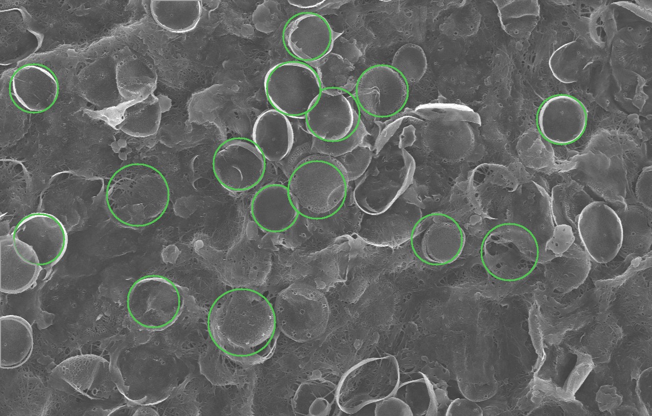
Fig. 2.1: Unedited output on a 3,000× image of Thalassiosira pseudonana. Each green circle is one of the 18 shells it found and measured.
Building a platform with W&M's Nano & Biomaterials Lab that automates diatom analysis in scanning-electron-microscope images.
Designed a pipeline that calibrates scale, segments shells with FastSAM, filters debris, and grades damage.
Measured 136 shells in one image in about 15 seconds on 2 CPUs, replacing one-at-a-time manual measurement.
Trained a random forest on 44 multi-scale texture features to identify samples, abstaining when unsure. It reached 77% accuracy on held-out imaging sessions (86% when confident), beating 47% and 59% baselines.
Validated the output: scale within 0.12% of the instrument's calibration, and median shell size inside the published range.
Deployed on Modal serverless behind a Cloudflare Worker, with a REST API, saved run history, and Excel export.